Figure 1. [The normal human retina fundus]. - Webvision - NCBI
Por um escritor misterioso
Last updated 22 dezembro 2024
![Figure 1. [The normal human retina fundus]. - Webvision - NCBI](https://www.ncbi.nlm.nih.gov/books/NBK554706/bin/Archetecture_Fovea-Image006.jpg)
The normal human retina fundus photo shows the optic nerve (right), blood vessels and the position of the fovea (center).
![Figure 1. [The normal human retina fundus]. - Webvision - NCBI](https://media.springernature.com/m685/springer-static/image/art%3A10.1007%2Fs11042-020-09041-y/MediaObjects/11042_2020_9041_Fig1_HTML.png)
Computerized retinal image analysis - a survey
![Figure 1. [The normal human retina fundus]. - Webvision - NCBI](https://www.ncbi.nlm.nih.gov/books/NBK554060/bin/466648_1_En_8_Fig1_HTML.jpg)
Fig. 8.1, [Cross-sectional OCT image of human retina with the corresponding cellular structures]. - High Resolution Imaging in Microscopy and Ophthalmology - NCBI Bookshelf
![Figure 1. [The normal human retina fundus]. - Webvision - NCBI](https://www.csbj.org/cms/attachment/126e4aa0-af36-45db-b0d5-914812fc7d76/gr1_lrg.jpg)
A review on automatic analysis techniques for color fundus photographs - Computational and Structural Biotechnology Journal
![Figure 1. [The normal human retina fundus]. - Webvision - NCBI](https://www.cell.com/cms/attachment/2119048143/2088495043/gr1.jpg)
Cell-Based Therapy for Degenerative Retinal Disease: Trends in Molecular Medicine
![Figure 1. [The normal human retina fundus]. - Webvision - NCBI](https://www.ncbi.nlm.nih.gov/books/NBK11556/bin/factsf5.gif)
Facts and Figures Concerning the Human Retina - Webvision - NCBI Bookshelf
![Figure 1. [The normal human retina fundus]. - Webvision - NCBI](https://www.ncbi.nlm.nih.gov/books/NBK11553/bin/clinicalergf24.jpg)
Figure 24, [Fundus photo and bright-flash ERG of patient with retinoschisis.]. - Webvision - NCBI Bookshelf
![Figure 1. [The normal human retina fundus]. - Webvision - NCBI](http://webvision.med.utah.edu/imageswv/huretina.jpeg)
Simple Anatomy of the Retina by Helga Kolb – Webvision
![Figure 1. [The normal human retina fundus]. - Webvision - NCBI](https://journals.physiology.org/cms/10.1152/physrev.00035.2019/asset/images/medium/z9j004202952r001.png)
Emerging Approaches for Restoration of Hearing and Vision
![Figure 1. [The normal human retina fundus]. - Webvision - NCBI](https://www.ncbi.nlm.nih.gov/corehtml/pmc/pmcgifs/bookshelf/thumbs/th-webvision-lrg.png)
Facts and Figures Concerning the Human Retina - Webvision - NCBI Bookshelf
![Figure 1. [The normal human retina fundus]. - Webvision - NCBI](http://webvision.instead-technologies.com/wp-content/uploads/2014/06/DONFig28a1-300x259.jpg)
11.2 The Electroretinogram and Electrooculogram: Clinical Applications. by Donnell Creel – Webvision
Recomendado para você
-
Retina - American Academy of Ophthalmology22 dezembro 2024
-
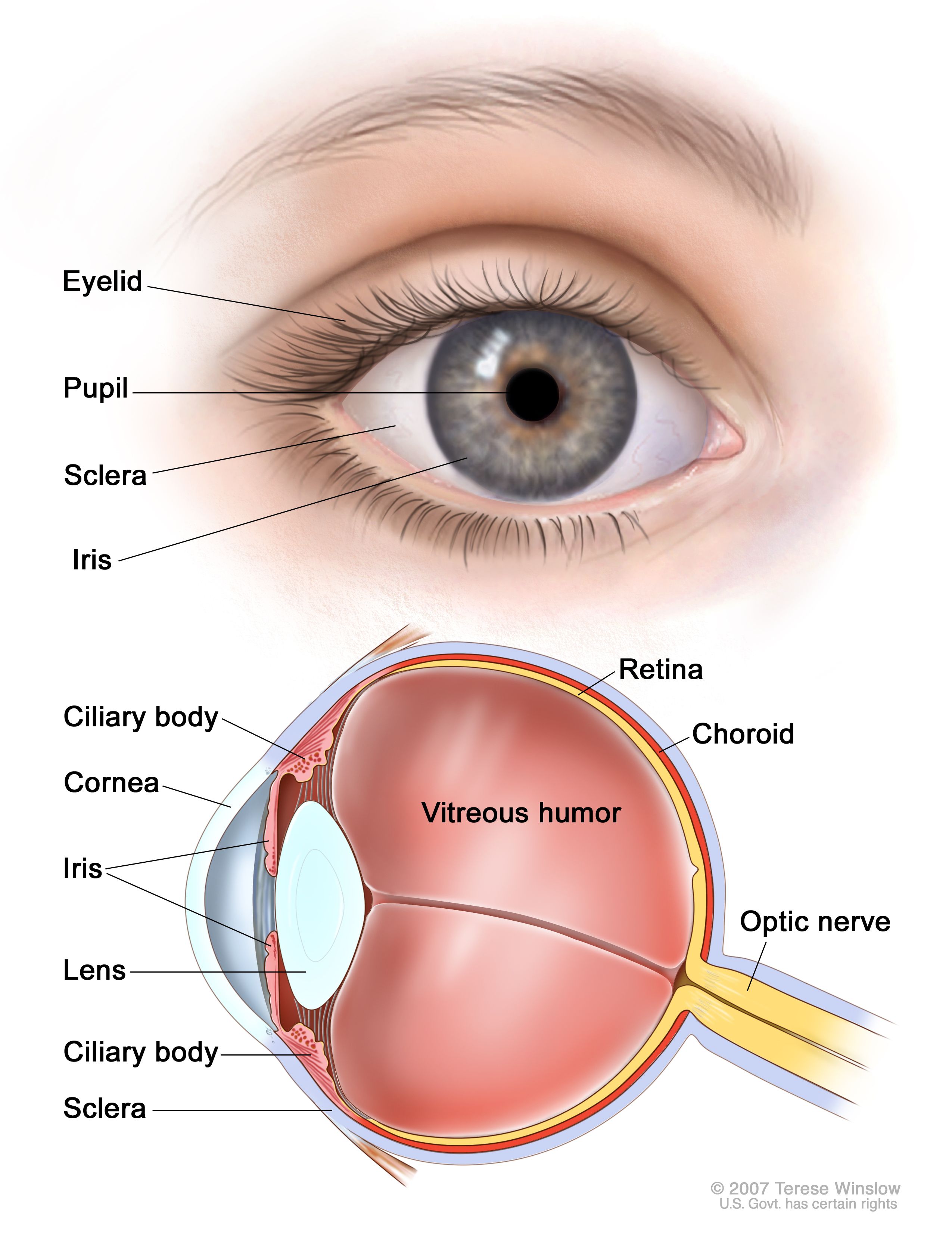 Retinoblastoma Treatment - NCI22 dezembro 2024
 -
 What Is a Detached Retina? - Outlook Eyecare22 dezembro 2024
 -
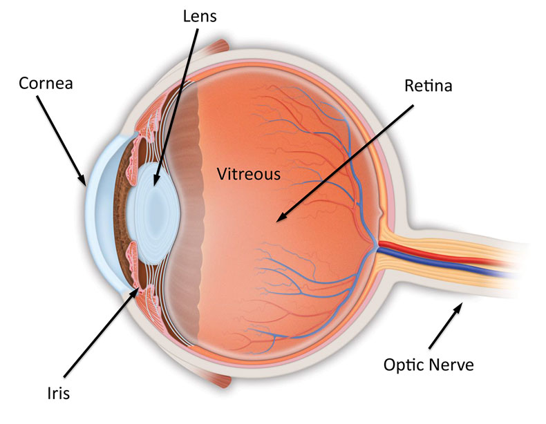 Anatomy of the Eye - Retina-Vitreous Surgeons of CNY22 dezembro 2024
 -
 Types of Retinal Detachment, Their Causes, and Treatments22 dezembro 2024
 -
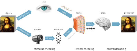 Research, Stanford Artificial Retina Project22 dezembro 2024
 -
 Retina Definition, Anatomy & Function - Video & Lesson22 dezembro 2024
 -
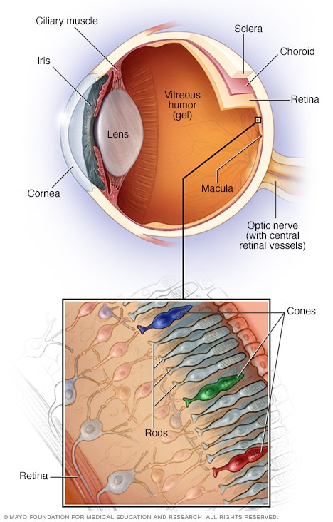 Retinal diseases - Symptoms and causes - Mayo Clinic22 dezembro 2024
 -
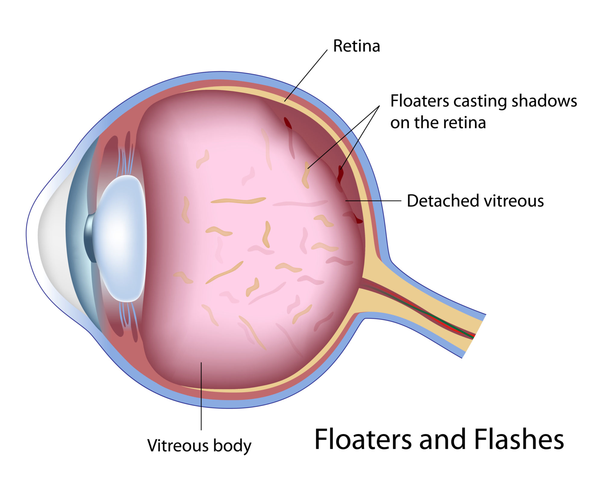 Vitreous Separation - Retina Vitreous Consultants, Inc22 dezembro 2024
 -
 Detached Retina, Optometrist in Chicago, Illinois22 dezembro 2024
você pode gostar
-
 Naruto – 1ª Temporada Completa (7 Discos)22 dezembro 2024
 -
 Rinkhals Snake Plays Dead, Deadly 6022 dezembro 2024
 -
 Boruto: Anime será dublado em português do Brasil22 dezembro 2024
 -
 Pulsefire Zed - KillerSkins22 dezembro 2024
 -
Pudim play22 dezembro 2024
-
 Freeze Dance Songs - Sing and Dance Along with THE KIBOOMERS - 15 Minutes22 dezembro 2024
 -
![Lost Ark Lost Ark Gold 50-100K [NA SERVERS] Fast Delivery](https://i.ebayimg.com/images/g/19kAAOSwbTxlC9yB/s-l1200.webp) Lost Ark Lost Ark Gold 50-100K [NA SERVERS] Fast Delivery22 dezembro 2024
Lost Ark Lost Ark Gold 50-100K [NA SERVERS] Fast Delivery22 dezembro 2024 -
 The Mountain: Into the Nether22 dezembro 2024
 -
 Mega Don Slot (Play N Go) Review + Demo22 dezembro 2024
 -
 KOS-MOS, Project X Zone Wiki22 dezembro 2024
